Pioneering microscopy for materials science

Microscopy has undergone significant advancements in recent years, transforming the way scientists investigate the microscopic world with unprecedented precision.
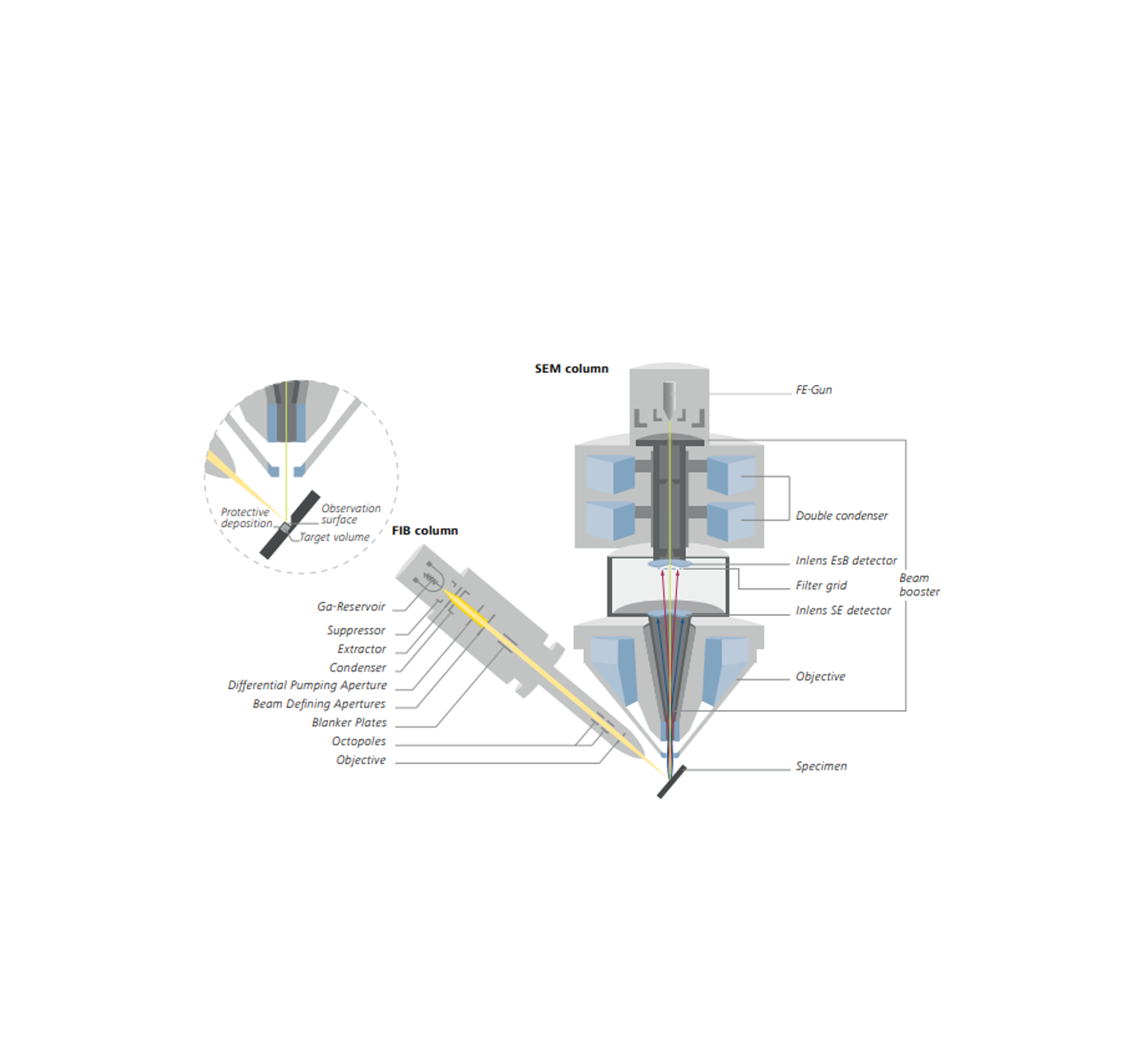
For instance, scanning electron microscopy (SEM) is a technique that employs a focused beam of electrons to produce high-resolution images of a sample's surface. Alternatively, focused ion beam-scanning electron microscopy (FIB-SEM) integrates SEM capabilities with the processing ability of a focused ion beam, enabling sample preparation and imaging of samples at nanoscale resolutions. This dual-beam approach has opened new avenues for studying materials with unparalleled precision.
3D Laser scanning microscopy (LSM) has also emerged as a versatile tool not only for imaging biological specimens but also for materials research with exceptional spatial resolution. By employing laser light in a confocal beam path, the microscope captures defined optical sections of the sample and combines them in a three-dimensional image stack. Together with fluorescence contrast an LSM allows scientists to investigate bio functionality of the specimen’s surface making it a valuable research tool for fields like cellular and molecular biology, or soft materials.
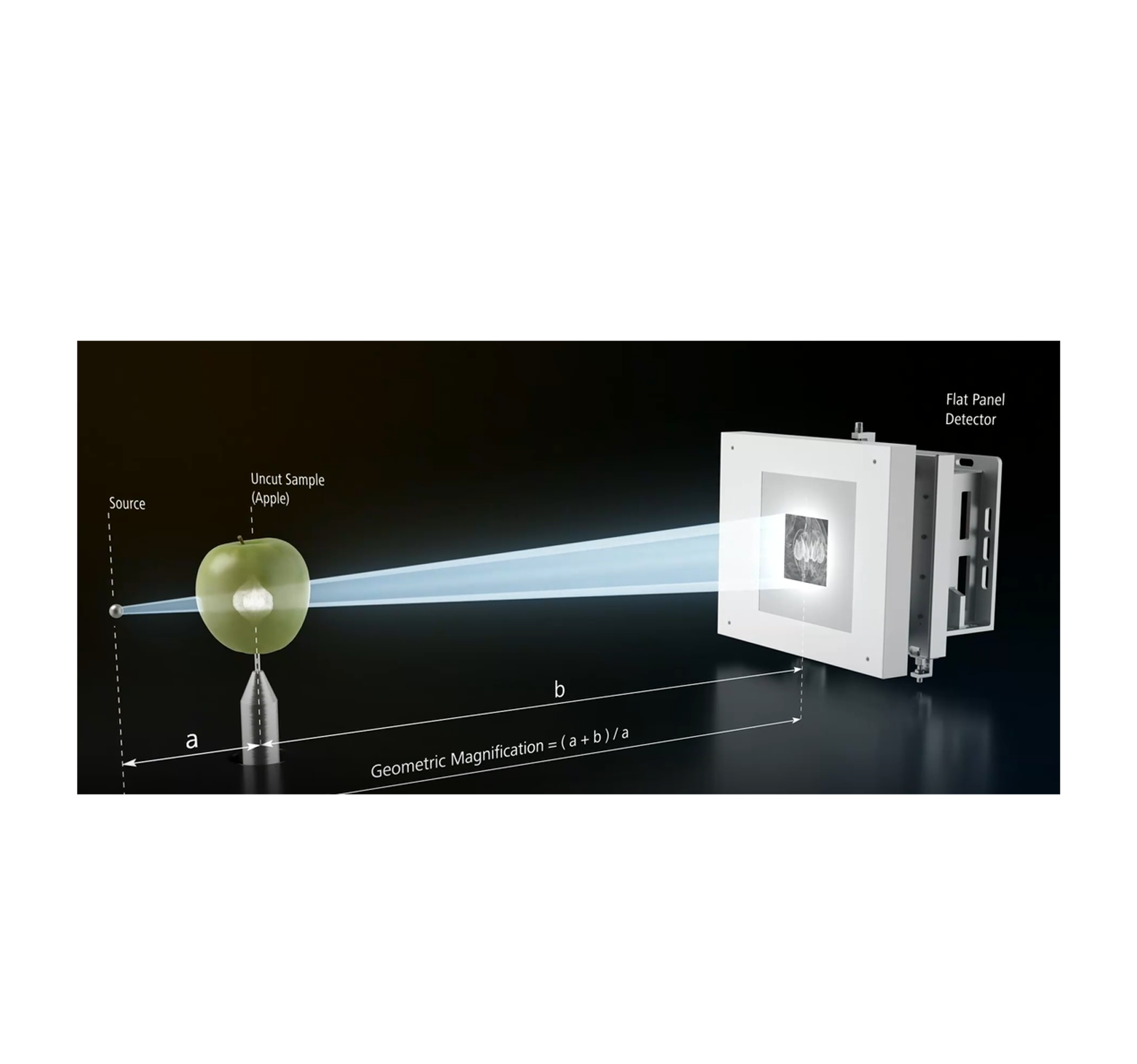
X-rays are not only used for imaging in computed tomography systems (CT) but also in genuine microscopes. With X-ray microscopy (XRM) users can examine properties and behaviors of their specimens non-destructively and in 3D. They can also develop and confirm models or visualize structural details. Submicron resolution at high contrast can be achieved even for relatively large samples by using lab-based synchrotron-like optics.


The company known for offering such tools is ZEISS, recognized for its reputation in delivering cutting-edge microscopy technologies that empower researchers to advance their studies. ZEISS’ dedication to advancing the field of microscopy is evident by its extensive range of instruments, which includes super-resolution microscopes, X-ray and scanning electron microscopes, and specialized software designed for correlation and AI-based image processing. These are particularly valuable when it comes to analyzing biomaterials, pharmaceutical substances, and polymers.
Within this comprehensive resource, explore the essential tools required for the characterization and investigation of various materials. From the analysis of soft materials and biomaterials to the characterization of polymers, gain access to a range of resources to advance your understanding of material science.

Soft materials
Soft materials lie in-between liquids and solids, showing characteristics that blend the two states. These materials typically possess low mechanical stiffness and are responsive and deformable under applied stress and external stimuli. Unlike solid materials, which maintain their shape and rigidity, soft materials can undergo significant changes in structure, making them adaptable for a wide range of applications.


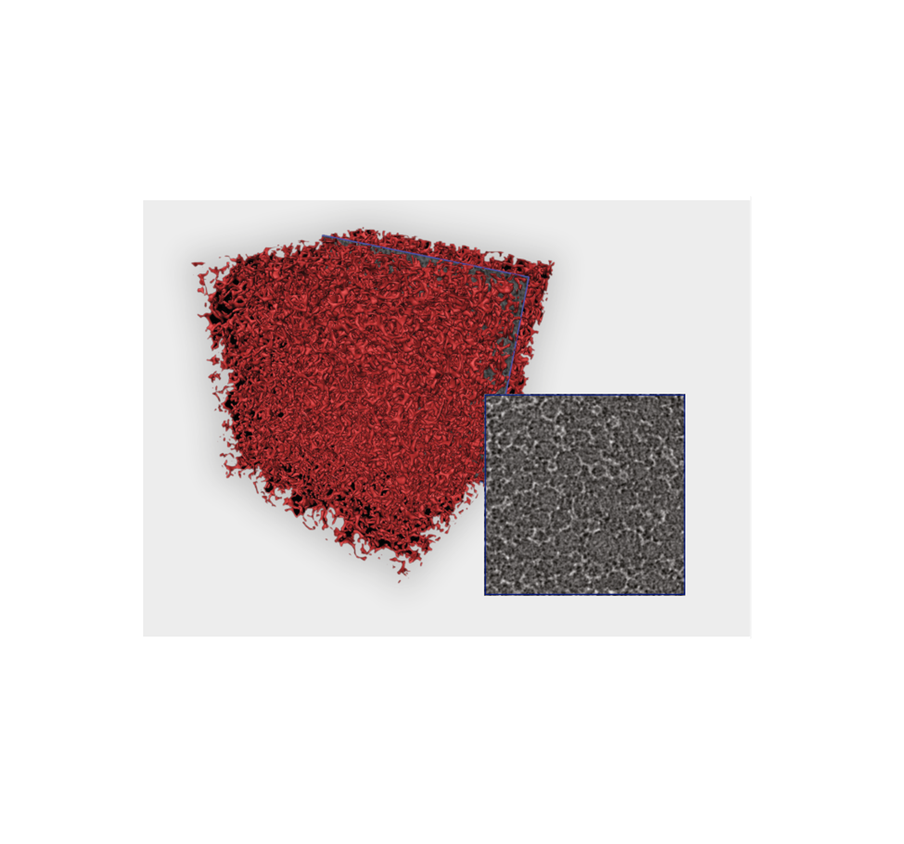
High-resolution Characterization of Solid Polymer Foams by X-ray microscopy, acquired with ZEISS Xradia Ultra
Examples and applications
Understanding the structure and behavior of soft materials is crucial for optimizing their performance and designing new materials with tailored properties.
Microscopy plays a key role in characterizing soft materials due to their complex structures. Traditional microscopy techniques, such as optical microscopy, provide valuable insights into the morphology of soft materials. However, to really access and study their complex internal structures and dynamics at the nanoscale, advanced microscopy techniques are often required. Transmission electron microscopy (TEM) and scanning electron microscopy (SEM) are is one indispensable tools for studying the structure of soft materials with nanometer resolution. For instance, TEM allows for the visualization of internal features by transmitting electrons through thin sections, while focused ion beam-scanning electron microscope (FIB-SEM) adds sample preparation to the range of capabilities. For the investigation of soft materials, cryo FIB-SEM is particularly interesting due to its ability to preserve and investigate samples in their near native state.
Explore the resources below to understand the critical role of studying consumer product microstructure in optimizing product development, facilitated by cryo-FIB-SEM techniques. Plus, gain insights on how to efficiently setup a ZEISS FIB-SEM system and explore 3D X-ray microscopes for solid foam characterization.
Integrating SEM and Raman imaging
Correlative Raman imaging scanning electron microscopy is a powerful analytical technique that combines the high-resolution imaging capabilities of SEM with the molecular specificity of Raman spectroscopy. By integrating these two powerful techniques, researchers can simultaneously visualize the morphology of samples at nanoscale resolution while obtaining detailed chemical information about their composition. This interdisciplinary approach holds great promise for advancing research in fields such as materials science, biology, and nanotechnology.
Explore the range of resources below to find out more about how to use this tool effectively in your research and discover how the Gemini electron optical design of ZEISS' field emission scanning electron microscope (FE-SEM) facilitates the examination of consumer product microstructures. Plus, watch an exclusive webinar with Parmesh Gajar, principal scientist at the University of Manchester and Darragh Murnane, Professor of Pharmaceutics at the University of Hertfordshire, as they discuss the characterization of asthma inhalation blends using correlative, AI powered X-ray microscopy.


Biomaterials
Biomaterials comprise of a diverse range of materials engineered to interact with biological systems for therapeutic or diagnostic purposes. These materials can range from natural sources like collagen to synthetic polymers and ceramics meticulously designed to mimic biological structures. Biomaterials play pivotal roles in medical implants, drug delivery systems, tissue engineering scaffolds, and diagnostic tools, with their properties tailored to promote biocompatibility, mechanical stability, and controlled release kinetics. As the field continues to evolve, biomaterials hold promise for addressing pressing healthcare challenges and driving innovations in regenerative medicine, personalized healthcare, and beyond.
Selecting the appropriate microscopy tools for characterizing biomaterials is crucial for obtaining accurate and meaningful data. For instance, high-resolution and magnification capabilities are essential for visualizing the complex structures and features of biomaterials at the micro- and nano-scale levels, aiding in understanding their morphology and surface characteristics. It is also important for users to consider compatibility with a given sample to ensure accurate characterization without compromising sample integrity.
There have been many advancements in the field, from developing bioinks for 3D bioprinting, fabricating nanomaterials for drug delivery, such as liposomes,and biomimetic materials designed to mimic the structure and function of natural tissues and organs.
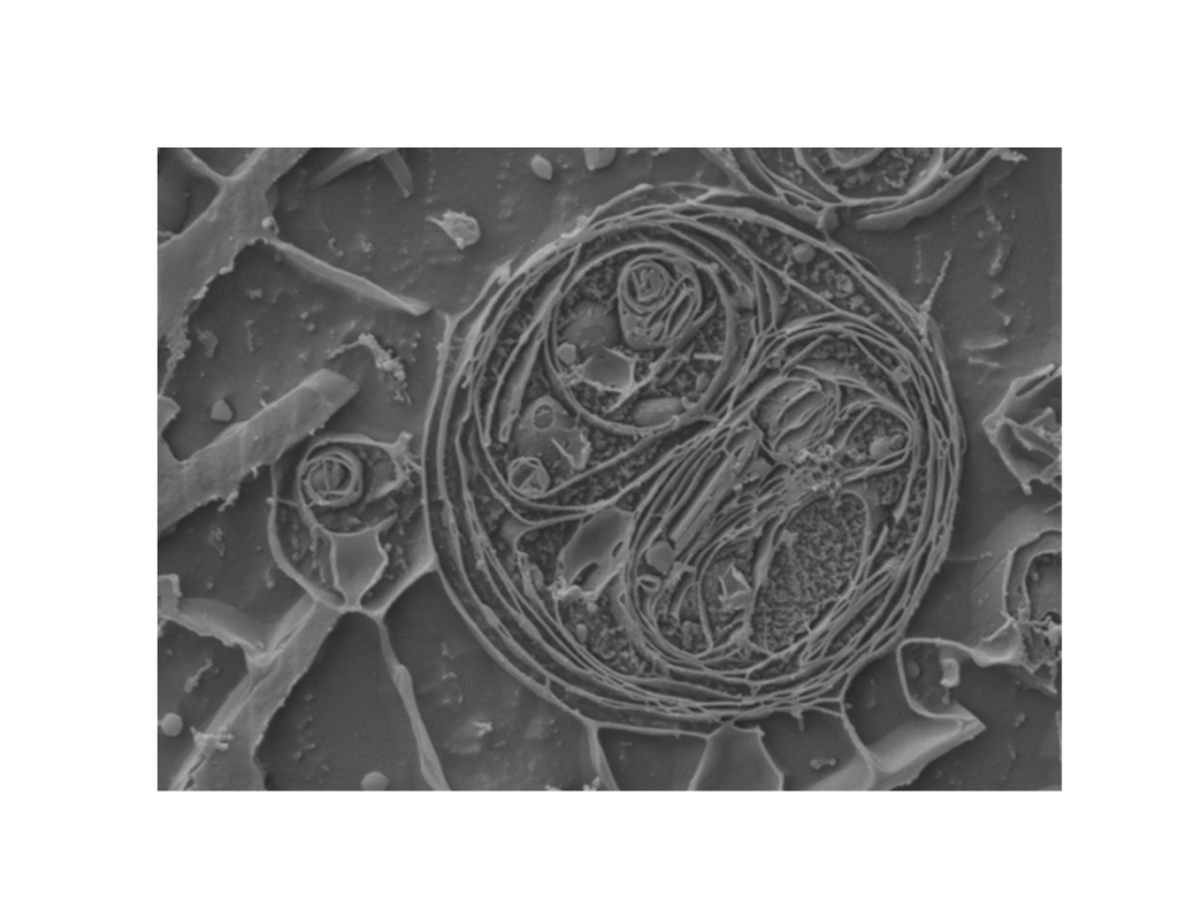
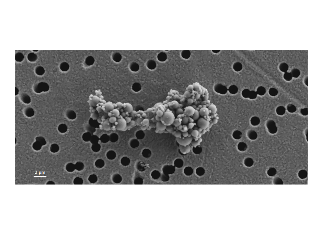
SEM image of a biomimetic surface for lotus effect, Sample courtesy: G. Umlauf, Fraunhofer IGB Stuttgart, DE

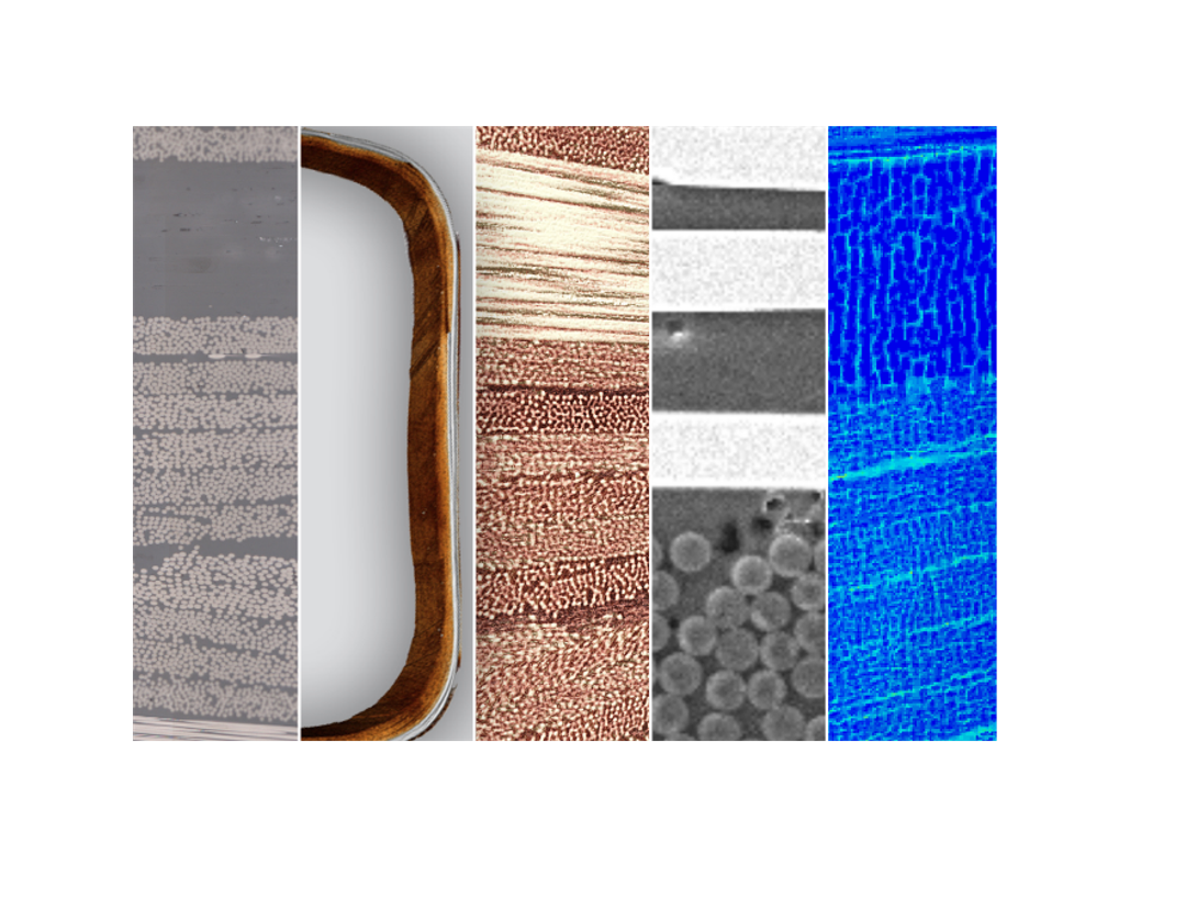
Distribution of fluorescent marked cells on a metal surface. Grey: titanium surface; green: cells; multi-channel analysis of surface structure, acquired with ZEISS LSM

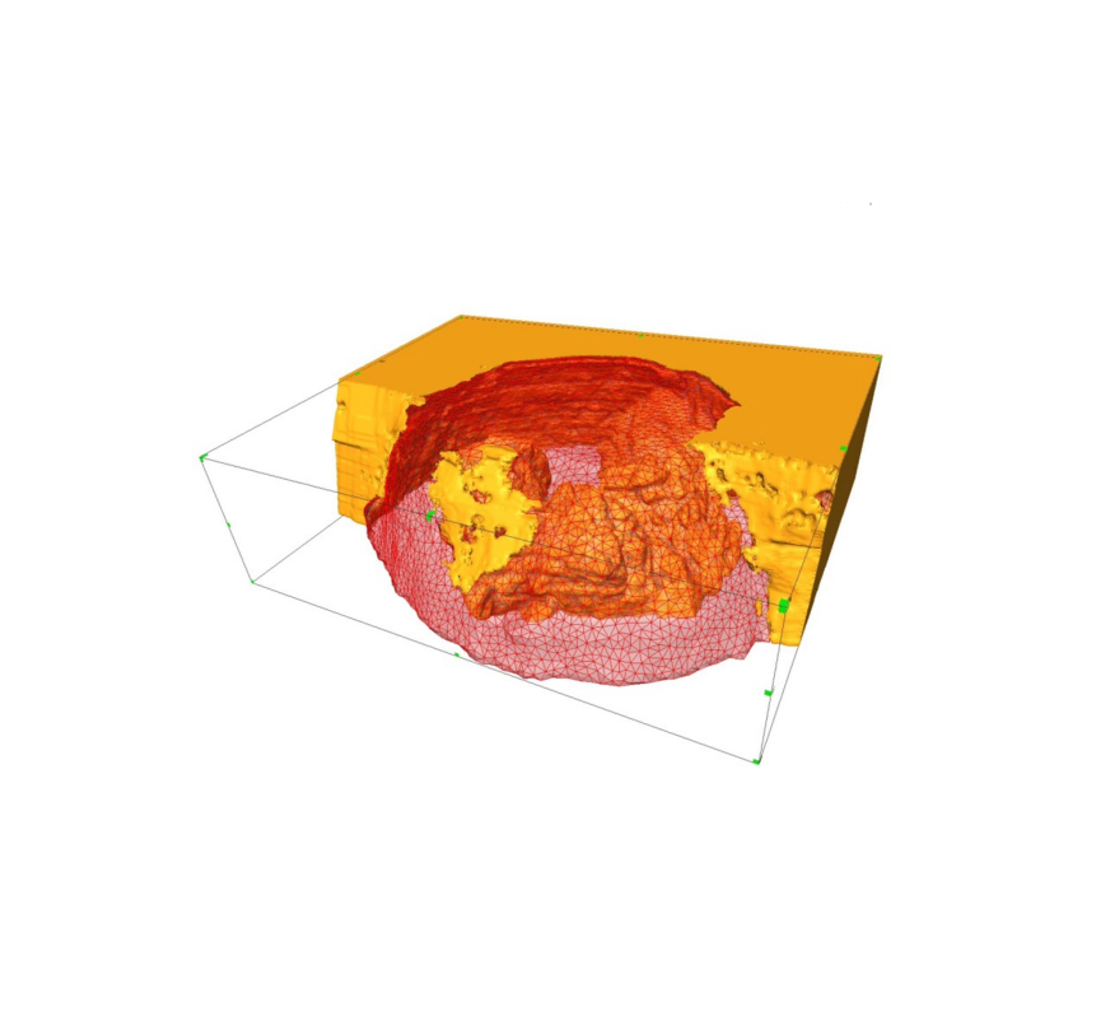
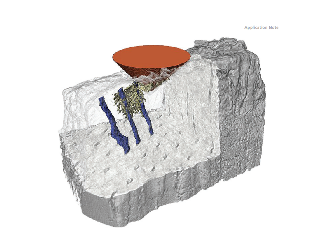
3D microstructure of scaffolds for tissue engineering, XRM dataset (ZEISS Xradia Ultra)
Explore the resources provided below for detailed insights into biomaterial characterization, including topics such as 3D imaging of crack growth in dentin and analysis of skin cream microstructure using advanced cryo-FIB-SEM technologies.

Polymers
Polymer characterization involves analyzing and understanding the structure, properties, and behavior of polymers, which are large molecules made up of repeating subunits called monomers. This process is crucial for various industries such as materials science, pharmaceuticals, and manufacturing, where polymers play a significant role. By characterizing polymers, researchers and engineers can optimize their properties for specific applications, ensure quality control during manufacturing processes, and develop new materials with tailored properties to meet desired performance requirements.
The accurate imaging of polymers plays a vital role in ensuring product quality, driving innovation, and advancing scientific knowledge in the field of polymer materials. Explore the resources below to find out more about how multimodal correlative imaging can be used to investigate microplastics, and the tools needed to better understand the structure-property relationships in a carbon-fiber composite.
Fractured surface of a polystyrene polymer, imaged with a ZEISS FE-SEM
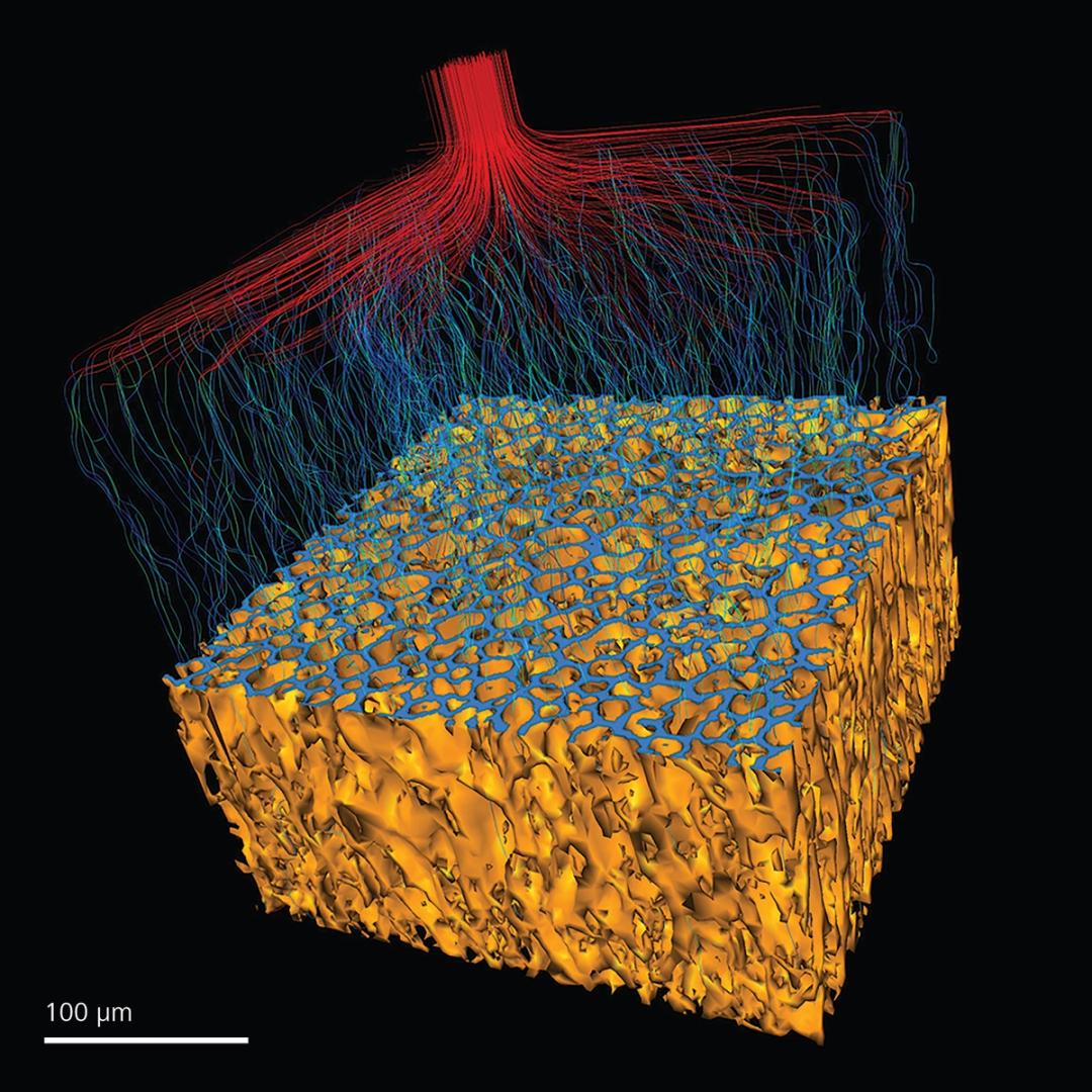
Porous polymer, 3D reconstruction of an XRM scan visualizing the flow of fluids, acquired with a ZEISS Versa XRM
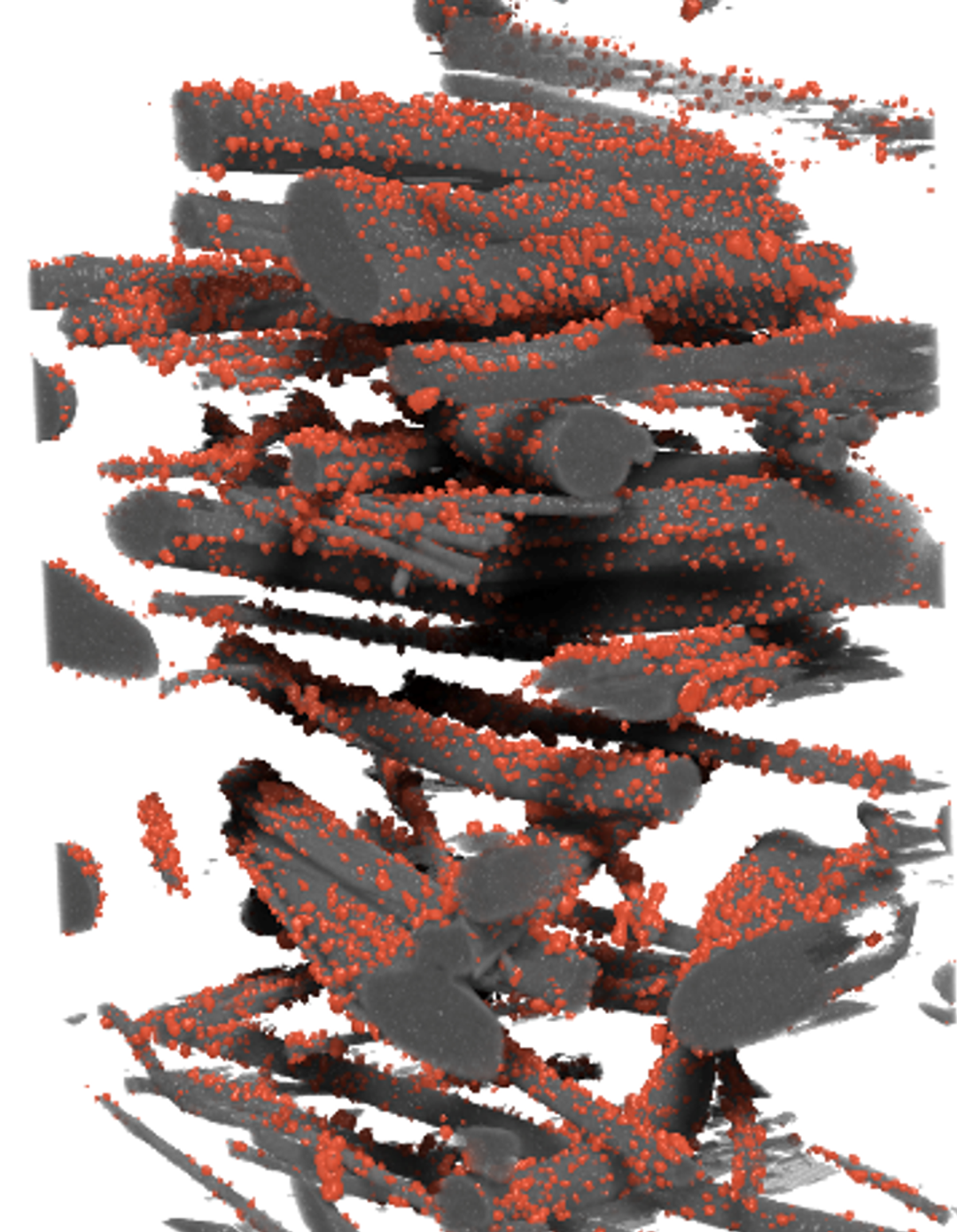
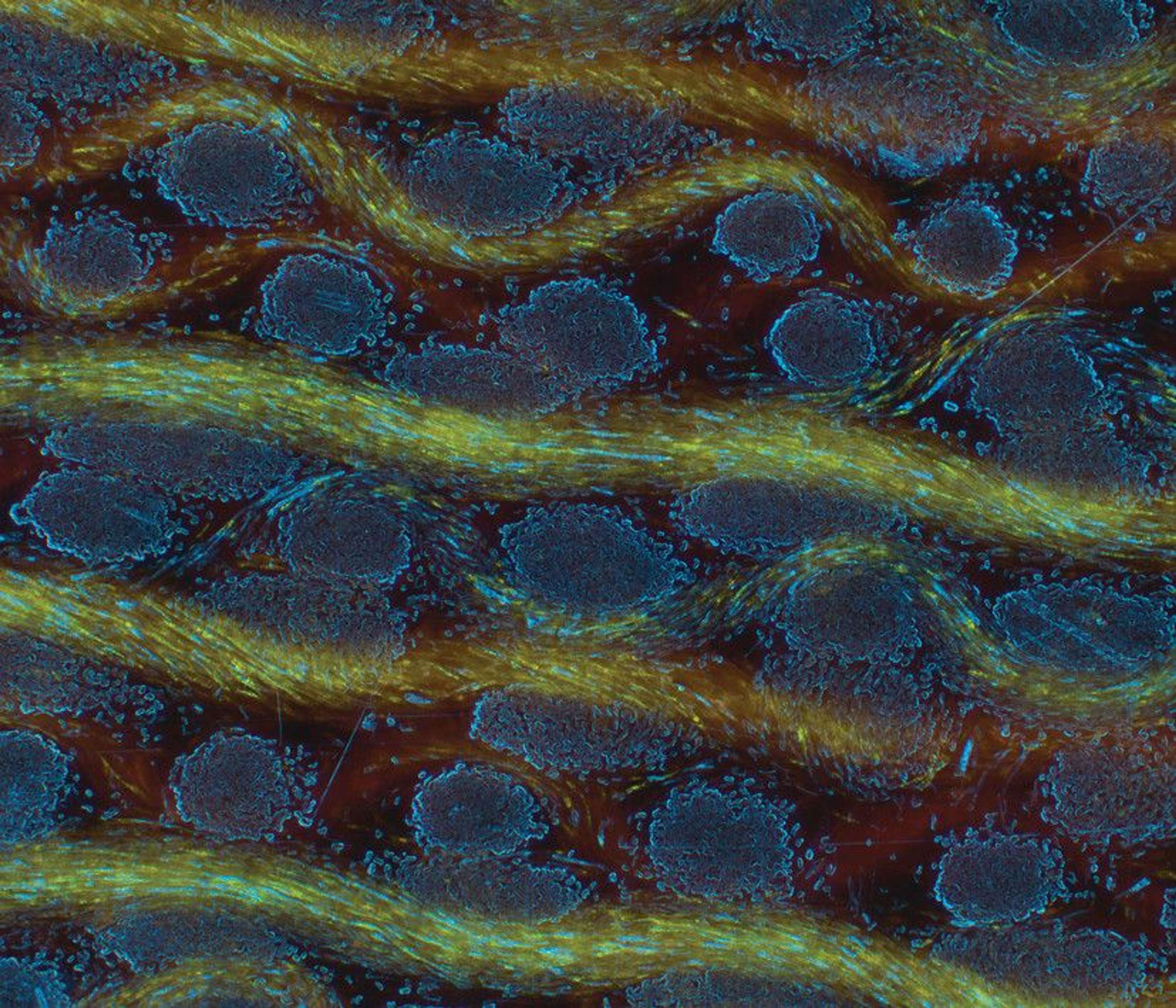
Polymer mask fibers with segmented NaCl particles to quantify filtering effectiveness. Imaged with ZEISS Xradia 810 Ultra
Technology

FE-SEM
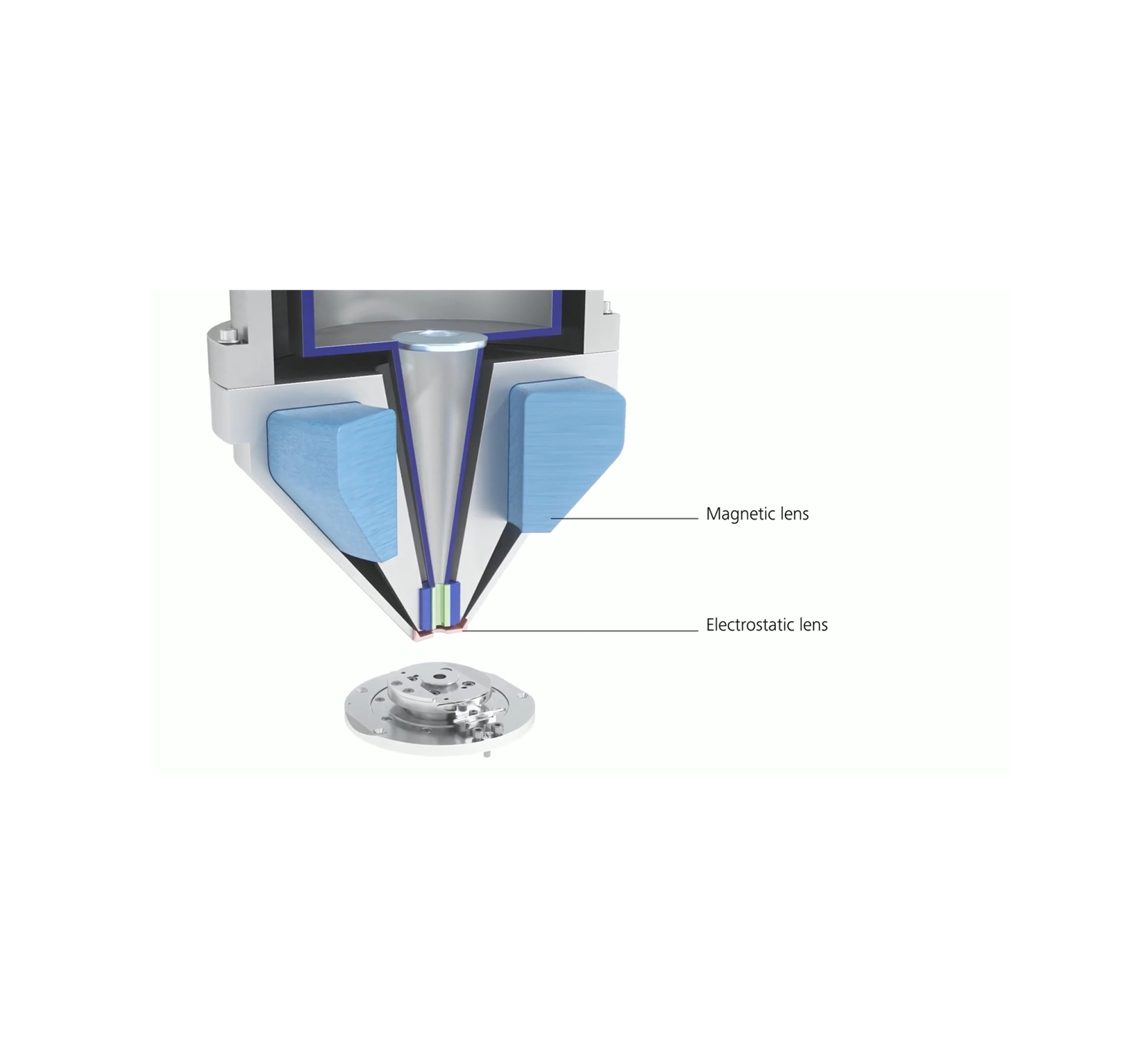
ZEISS GeminiSEM stands for effortless imaging with sub-nanometer resolution. Use it for your most demanding projects in materials and life science.
Request pricingLearn more

FIB-SEM (Crossbeam)
“Great product, essential to all our products; it never stands still in our lab!”
Silke Christiansen, Inam

ZEISS Xradia Versa X-ray Microscopes
“High throughput with good image quality.”
Arulraj Ramalingam, National University of Singapore
Request pricingLearn more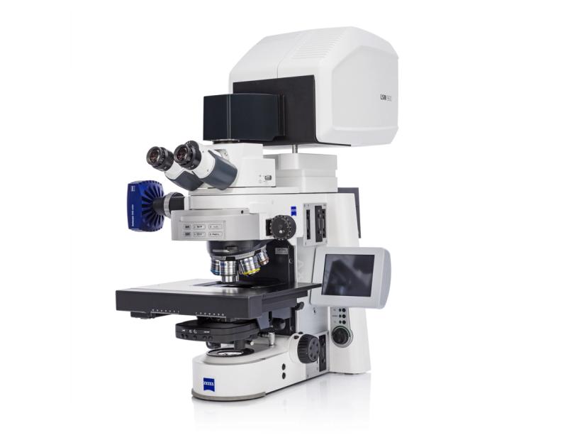
ZEISS LSM 900
“Surface analysis in a fast and deep way”
Jacopo Barberi, Politecnico Di Torino

ZEN core
“It is the best experience I have ever had with the ZEISS optical microscope in our fiber characterization Lab.”
Elif YAPAR YILDIRIM, Bilkent University UNAM
Request pricingLearn more