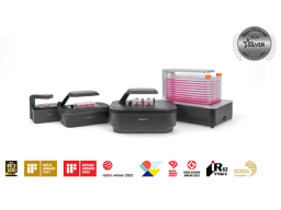
Celloger®, Automated Live Cell Imaging System from Curiosis
Capture the moments of dynamic cellular processes, improving your research with comprehensive insights


Receive your quote directly from the manufacturer.
Z-stacking function was a strong feature, allowing the acquisition of images at multiple focal planes
Biochemistry
For COS7 cell research, the Z-stacking function was a strong feature, allowing the acquisition of images at multiple focal planes and combining them to generate enhanced 2D images with improved depth information. The image stitching function was also valuable, facilitating the analysis of large cell populations by merging multiple images into a single comprehensive view.
Review Date: 16 Apr 2025 | CURIOSIS
Compatibility with various culture vessels was one of the most appreciated features
Biochemistry
In conjunctiva cell research, compatibility with various culture vessels was one of the most appreciated features. The system supported flasks, dishes, and well plates, allowing for flexible experimental setups. The user-friendly software made it easy to track cell confluency, monitor growth curves, and perform data analysis. The time-lapse video function was especially useful for visualizing dynamic cellular changes over time, making it an excellent tool for cell behavior analysis.
Review Date: 16 Apr 2025 | CURIOSIS
One of the most satisfying aspects of using Celloger® Mini Plus was its real-time monitoring capability
Biochemistry
One of the most satisfying aspects of using Celloger® Mini Plus for pterygium cell research was its real-time monitoring capability. It allowed continuous observation of cell growth and changes without disturbing the culture environment, ensuring stable data acquisition. Additionally, the multi-position imaging function enabled the simultaneous analysis of multiple samples, significantly improving experimental efficiency. The autofocus function was also highly reliable, providing consistently sharp images without losing focus.
Review Date: 16 Apr 2025 | CURIOSIS
Easy to use, quick running time
Organoid research
Easy to use, quick running time. Providing free analysis software would be beneficial.
Review Date: 16 Apr 2025 | CURIOSIS
The device is highly optimized for 2D cell imaging but also demonstrated promising capabilities in imaging organoids
Organoid research
The device is highly optimized for 2D cell imaging and, while originally designed for this purpose, it also demonstrated promising capabilities in imaging organoids with 3D structures. 1. High resolution and efficient imaging intervals The system enabled detailed observation of cellular structures with high resolution. Even when using multiple fluorescence channels, live imaging was possible at intervals as short as 30 minutes, which was extremely useful for time-lapse experiments. 2. Stability during imaging Imaging remained stable even when the media was changed or the plate was temporarily moved during the experiment. The transitions were also captured naturally in the resulting video. 3. Natural imaging of organoid growth Although there were some limitations, it was still possible to acquire relatively natural organoid growth images when specific organoids were properly targeted. This allowed us to collect data that effectively aligned with our research objectives.
Review Date: 16 Apr 2025 | CURIOSIS
It was easy to observe changes in both cells and GFP expression over time during the culture period
Cell culture
It was easy to observe changes in both cells and GFP expression over time during the culture period.
Review Date: 16 Apr 2025 | CURIOSIS
The system was very effective for checking transfection efficiency and expression levels in real time
Cell monitoring
In terms of performance, the system was very effective for checking transfection efficiency and expression levels in real time. However, the option to use higher magnification would be a valuable improvement.
Review Date: 16 Apr 2025 | CURIOSIS
It was very helpful to monitor the growth and wound healing capabilities of cells in real time.
Arthritis treatment and joint regeneration
It was very helpful to monitor the growth and wound healing capabilities of cells in real time. The automated time-lapse imaging system was also convenient and easy to use.
Review Date: 16 Apr 2025 | CURIOSIS
It offers clear fluorescence imaging without need for post-processing
Cancer cell study
Using this system, the fluorescence (GFP) is clearly visible in the images without the need for post-processing. It offers the convenience of setting the center position for all wells at once.
Review Date: 21 Oct 2024 | CURIOSIS
The system provides real-time bright field and fluorescence measurement and captures consecutive images for video
Drug delivery system
Real-time measurement of both bright field and fluorescence is possible inside the incubator. It is highly advantageous that consecutive images can be captured at set positions and later compiled into a video.
Review Date: 21 Oct 2024 | CURIOSIS
Key features of Celloger®
- - Real-time cell monitoring inside an incubator
- - Compatible with different vessel types
- - Time-lapse imaging capability
- - User-friendly functions included in the software package
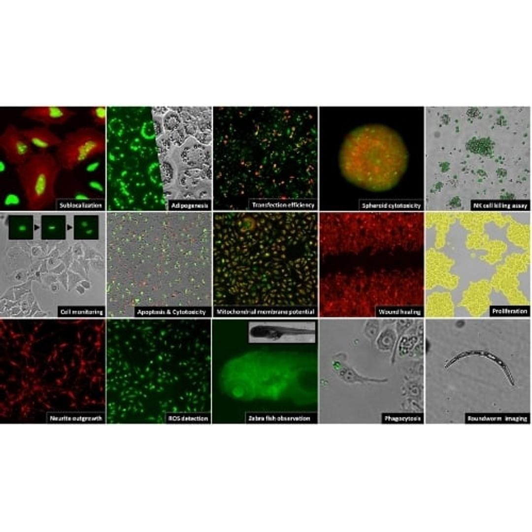
Key applications of Celloger®
- - Cell proliferation
- - Wound healing assay
- - Co-culture monitoring
- - Spheroid cell death assay
- - Actin dynamics assay
- - Mitochondrial membrane potential
- - ROS detection
- - Phagocytosis monitoring
- - Zebrafish observation


Product Overview
Links
Related Products
Request Quote for All Products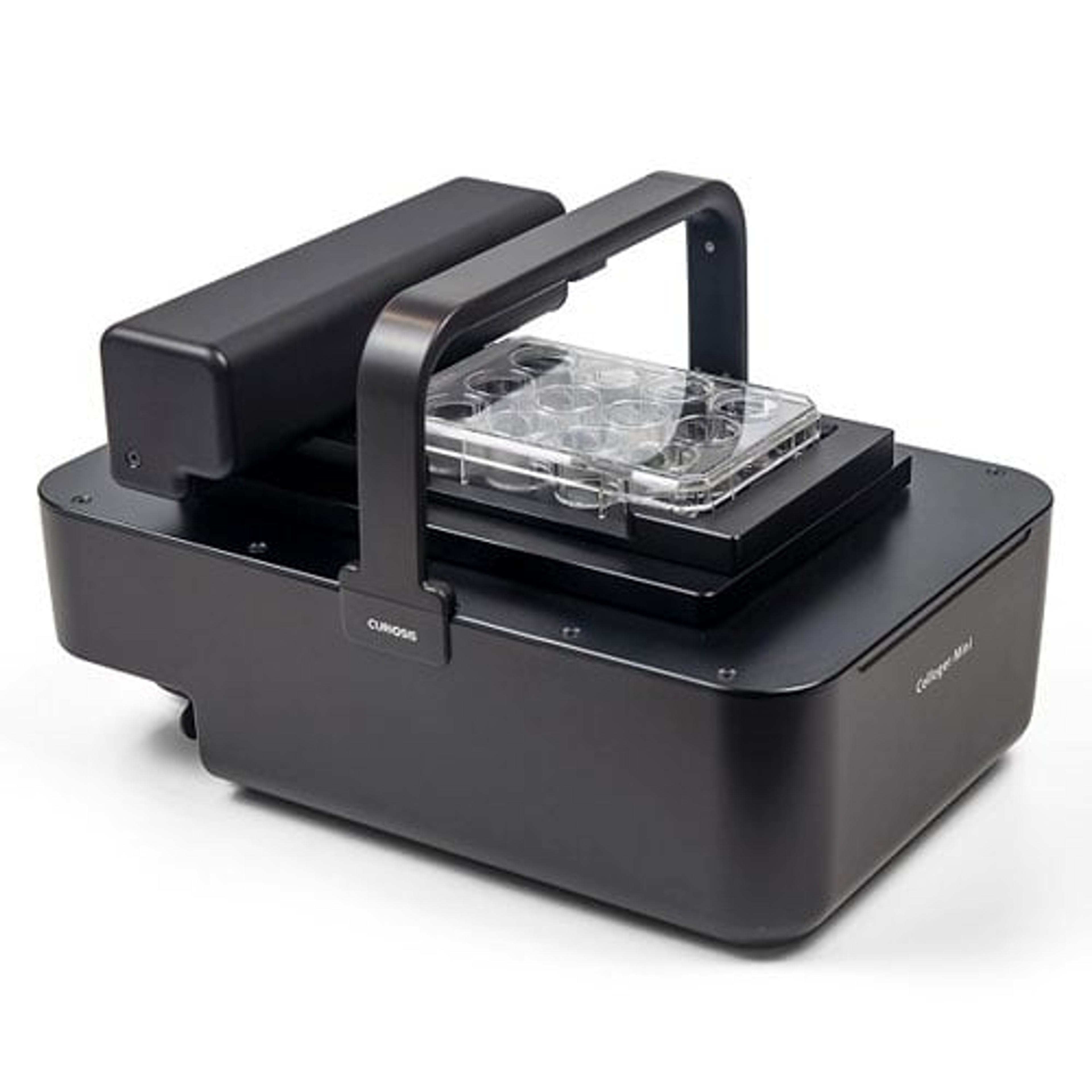
Products Model Information
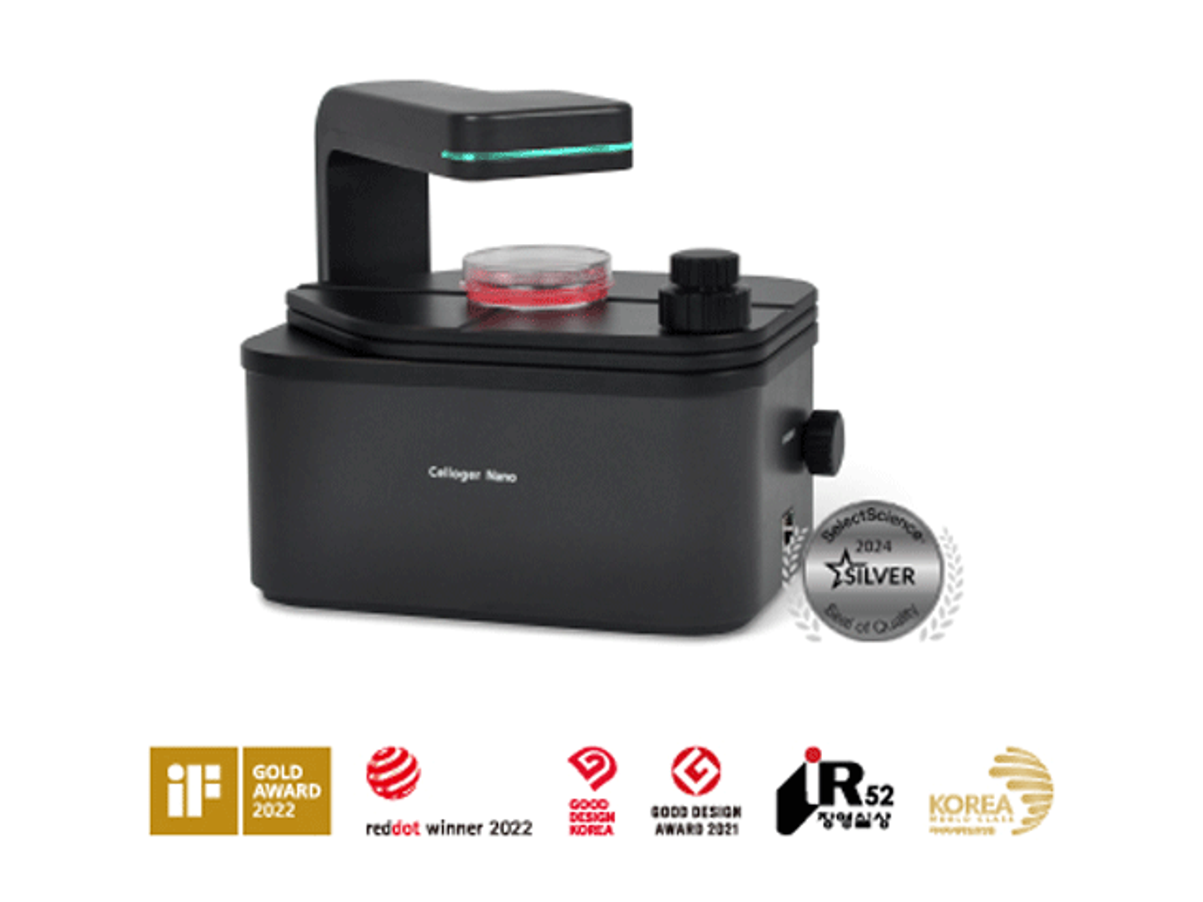
Celloger® Nano, Benchtop Digital Microscope from Curiosis
CURIOSISCelloger® Nano is a benchtop digital microscope equipped with a wireless connection, enabling you to check the state of your cells in real-time from any location within your laboratory. With all the necessary functions to check the cells, you can quickly assess the condition of your cells.
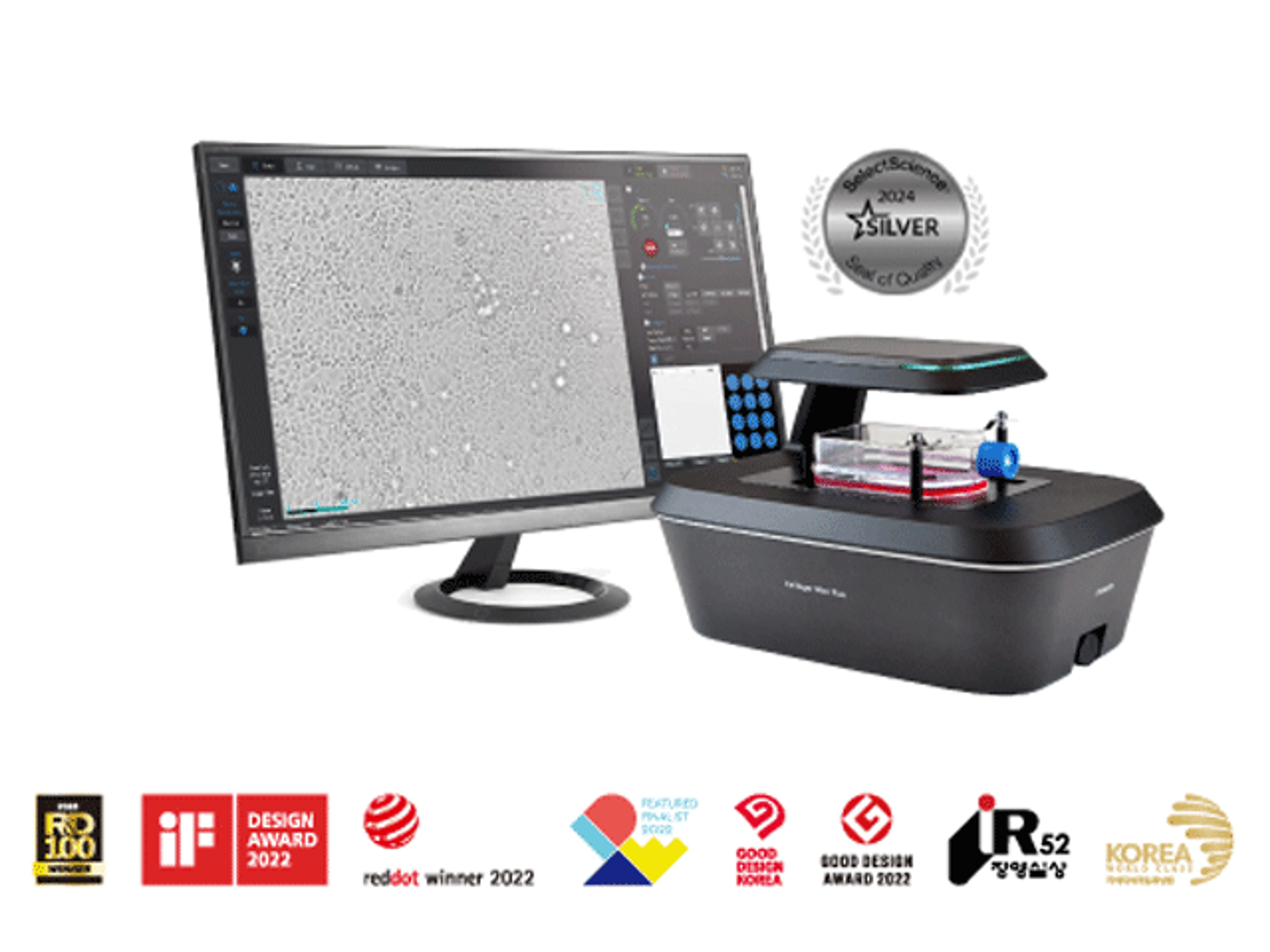
Celloger® Mini Plus, Automated Live Cell Imaging System from Curiosis
CURIOSISCelloger® Mini Plus is an automated live cell imaging system with fluorescence and brightfield microscopy. Celloger® Mini Plus makes it faster and easier to accumulate outstanding research results tailored to your research protocol.
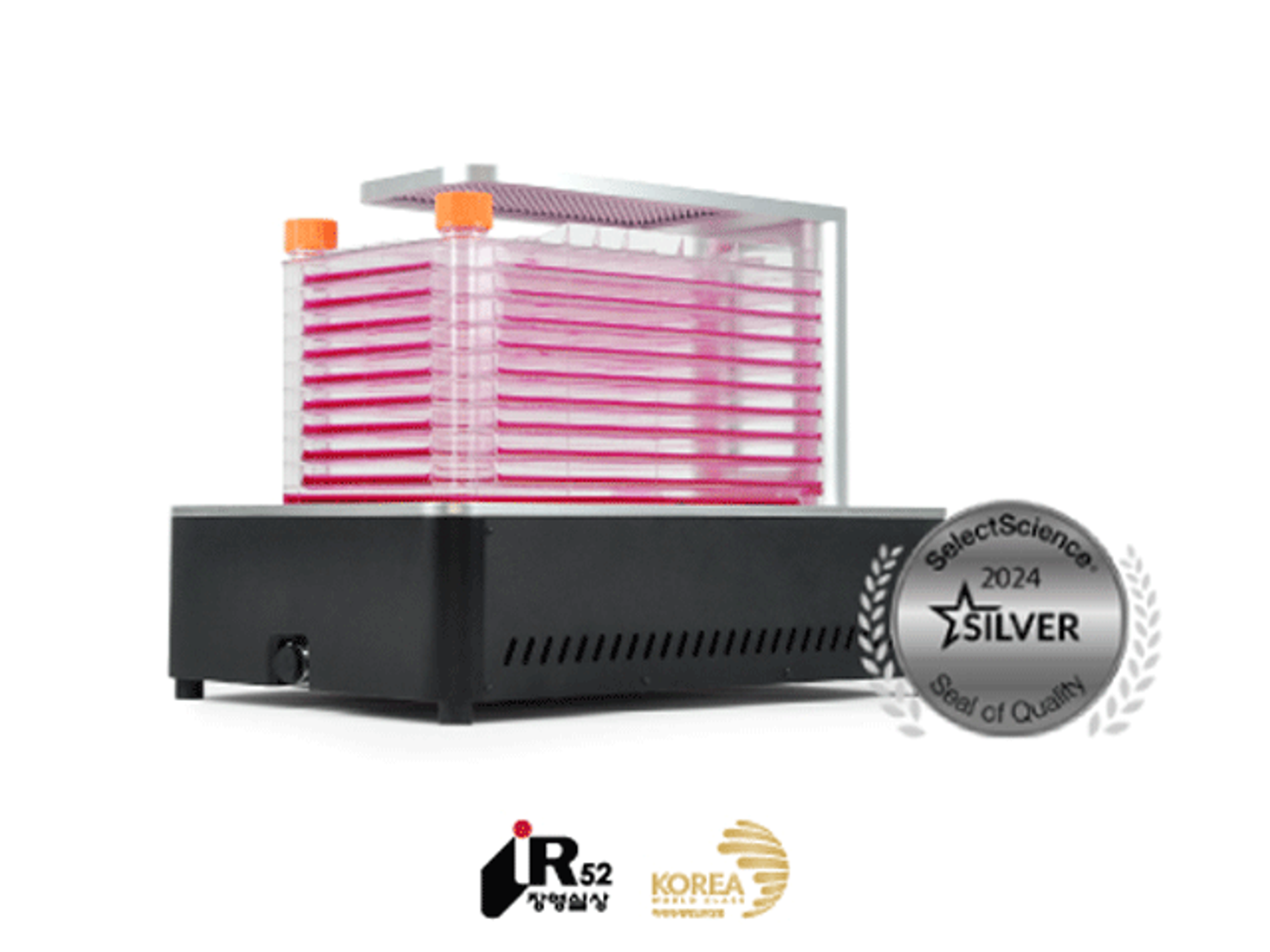
Celloger® Stack, Automated Multi-layer Vessel Monitoring System from Curiosis
CURIOSISCelloger® Stack is an automated multi-layer vessel monitoring system that enables real-time imaging of cell samples while they are cultured in an incubator.
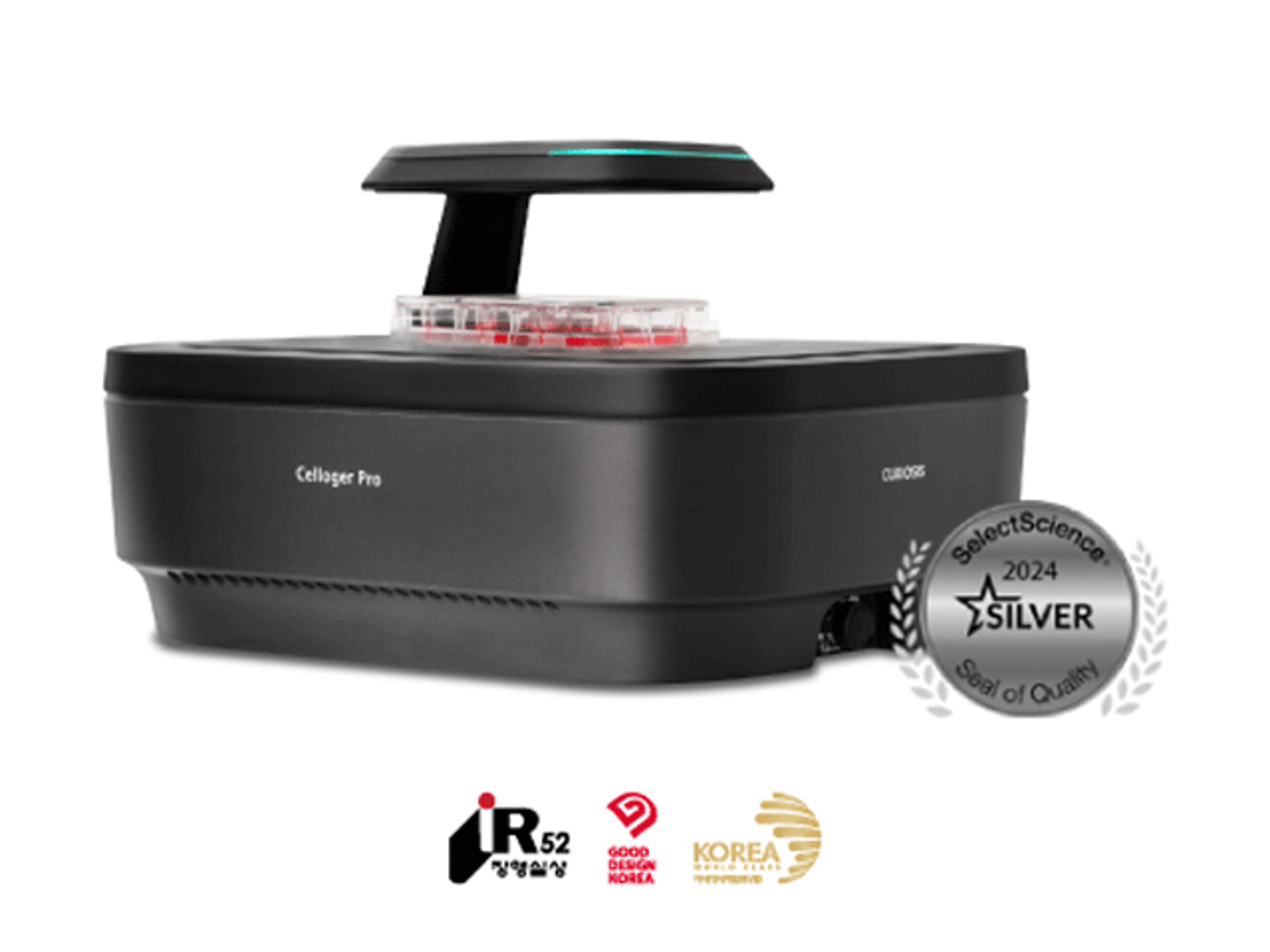
Celloger® Pro, Automated Live Cell Imaging System from Curiosis
CURIOSISWith its streamlined workflow and versatile capabilities, Celloger® Pro is a valuable tool for researchers seeking efficient, accurate, and cost-effective cellular analysis.